Raik's Page

Home Links Research LogoSwitch That's me Contact

Hacking Trypsin

Details of my Diploma Project

Here are some details about my diploma project, mainly based on the talk I gave on June 25th. Ok, this is after hours work so, please, ignore any mistakes.

Content
 
go to this section
1. Introduction
go to this section
2. Experimental Methods
2.1 Cloning and Expression of Trypsinogen Variants
2.2 Isolation of Trypsin Variants
2.3 Kinetic characterisation
go to this section
3. Docking Studies
3.1 Objective
3.2 Mechanism (Simulated Annealing)
3.3 Genetic Algorithm
3.4 Steps of a Docking Study
go to this section
4. Results
4.1 Peptide Synthesis with Model Peptides
4.2 Docking Experiments with Model Peptides
4.3 Conclusions
go to this section
5. References

go to top
1. Introduction
go to previous section  go to next section

Based on their catalytic mechanism it is possible to employ serin proteases like trypsin for enzymatic peptide bond formation. Especially, substrate mimetics - a new class of artificial substrates, allow for efficient ligations of peptides and amino acids, as demonstrated in recent years.
However, if the product contains specific cleavage sites recognised by the protease, this ligation is counteracted by the (natural) proteolytic reaction.

A promising approach to overcome this problem is the manipulation (i.e. eradication)of the enzyme's natural specifity.

Figure 1a shows the 3-dimensional structure of rat trypsin with a bound substrate, in this case Cys-Lys-Ala - the central residues of the soy-bean trypsin inhibitor (based on pdb structure 3tgi).
Note, how the side chain of Lys is buried within the S1 binding pocket of the enzyme.

Click on the image to view a larger version.

Fig. 1a: wild-type rat trypsin.

Figure 1b gives a more detailed insight into the catalytic center and the S1 binding pocket of rat trypsin, based on the same structure as Figure 1a.
This time, some of the protein's residues are shown, as well: the residues of the catalytic triade (D102, H57, S195, G104) in light blue, D189 and S190 in green.
The latter two are highly important for the enzyme's specifity. Being situated at the bottom of the binding pocket, they stabilise the binding of the substrate by forming a salt bridge and a hydrogen bond with the positively charged side chain of Arg or Lys.

Click on the image to view a larger version.

Fig. 1b: trypsin, catalytic center

It has been shown previously that alterations within this binding pocket, first of all, affect the amidase activity of trypsin. By contrast, the esterase activity (which is needed for peptide synthesis reactions) often can be retained. Building upon this, I prepared and isolated two variants of trypsin with altered S1 binding pocket: trypsin D189A and trypsin D189A,S190A.
These variants should no longer accept Arg or Lys as substrates, whereas some substrate mimetics should still be utilised. Hence, the new enzyme would be suited to perform peptide ligations without the limitations raised by proteolysis.
To validate this assumption the proteins have been characterised by HPLC-based acyl-transfer experiments.

An important prerequisite of this project is detailed knowledge about the enzyme's structure and function at an atomic level. Modern software tools based on molecular mechanics allow for investigations going well beyond the classical "look and speculate" approach:
The AutoDock software package makes it possible to simulate the binding of many substrates to the altered enzyme in silico. Docking studies were therefore part of this project (see section 3 for details) and yielded the structural basis upon which to interpret the experimental results.

go to top
2. Experimental Methods
go to previous section  go to next section

2.1 Cloning and Expression of Trypsinogen Variants

Starting point for the site directed mutagenesis of trypsin was the E. coli plasmid pST. pST is based on pBluescript and contains the wild type rat trypsinogen gene situated next to a yeast ADH promoter and and an alpha leader factor. After completing the necessary manipulations in E. coli, the insert containing the altered trypsinogen gene was transfered into the yeast shuttle vector pYT.
Yeast cells, transformed with the new constructs, secreted the recombinant proteins into the culture medium.

2.2 Isolation of Trypsin Variants

The trypsinogen was purified from the culture supernatant by cation exchange chromatography and cleaved with enteropeptidase to obtain the mature protein. Perfusion chromatography was then applied for further purification.

2.3 Kinetic Characterisation

Model reactions of the type
AAXAG + G -----> GAAXAG

whith X being Arg, Lys, Phe or Tyr have been performed in order to test the new variants' suitability for peptide ligations. Gly was applied as substrate mimetic coupled to guanidinophenol (Bz-Gly-OGP). At regular time intervalls, aliquotes of the reaction mixture were withdrawn and analysed by HPLC.

go to top
3. Docking Studies
go to previous section  go to next section

3.1 Objective

Docking studies with AutoDock predict the bound conformations of flexible ligands to rigid macromolecules. Apart from this structural data, the interaction energy upon binding (and therefore the binding constant)is estimated based on an empirical free energy function.

3.2 Mechanism: Simulated Annealing

One simulation of ligand binding is called docking run. Each docking run consists of repeated variations of the ligand's geometry (and position) followed by an energy evaluation for the whole system of protein and ligand (based on a force field related to AMBER). In order to make this process computationally tractable, the program applies pre-calculated energy grids serving as lookup tables during the docking run.

3.3 New Mechanism: Genetic Algorithm

In contrast to the Monte Carlo simulated annealing search described in the previous image, the new version of AutoDock uses a genetic algorithm to find energetically optimal ligand conformations. This method proved to be more efficient and reliable.

3.4 Steps of a Docking Study

1. preparation of ligand structures
2. preparation of protein structures
3. calculation of energy grids
4. several automated docking runs (10-20)
5. evaluation of results

go to top
4. Results
go to previous section  go to next section

Below are some selected results of the project. (I'm not going to present my whole thesis here...)

4.1 Peptide Synthesis with Model Peptides

The possibility to use the new trypsin variants for peptide synthesis has been demonstrated in model reactions by transferring Gly to peptides containing natural cleavage sites of trypsin and chymotrypsin.

Synthesis of a peptide containing lysine
As shown in figure 4.1a, trypsin D189A/190A is feasible for synthesising peptides that contain Lys or Arg (note the product concentration reaching a yield of nearly 80%). With variant D189A the situation is somewhat different: The enzyme still exhibites a (reduced) specifity towards Lys- or Arg-containing peptides. However, in contrast to wild type Trypsin (preferring Arg), Lys is the preferred substrate.

Both enzyme variants can be employed for synthesis of Phe- or Tyr-containing peptides. It must be noted, however, that the synthesis of Tyr-containing peptides is limited by a novel chymotrypsin-like specifity. Although somewhat disappointing, this came not entirely surprising for trypsin D189A, especially taking into account the published data about trypsin D189S. At first unexpected, however, was the finding that trypsin D189A/S190A too, is exhibiting a significant (even slightly higher) specifity towards tyrosine substrates. By contrast S190 was believed to mediate tyrosin specifity by interacting with the p-OH group of tyrosin substrates. According to this, replacing S189 by Ala should, in fact, impair any chymoptrypsin-like specifity.
A reasonable explanation for this effect could be gathered from the docking experiments.

4.2 Docking Experiments with Model Peptides

Initially, docking studies (combined with experimental examinations) have been employed to assist the search for an optimal substrate mimetic. Since the results obtained proved to be useful for explaining the enzymes new specifities, the studies were later extended to model peptides containing Arg, Lys, Tyr or Phe. An example of the latter results is presented here.

Docking of a Lys-containing peptide to wt-Trypsin

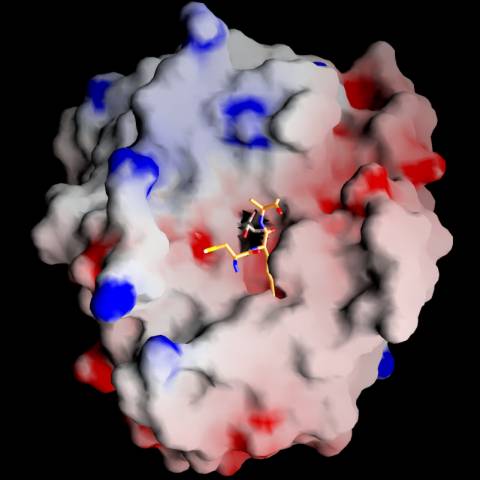
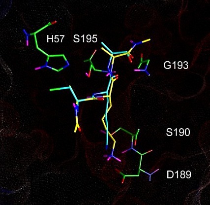
Figure 4.2a shows the docked arrangement of Ac-Ala-Lys-Ala-Me bound to wt-trypsin. The lowest energy structure found (yellow) resembles the arrangement of SBTI's central residues Cys-Lys-Ala (blue, the same as shown in Fig. 1a) in the pdb structure 3tgi with an RMSD of only 0,66 A². This result is a good confirmation of the accuratness of docking studies and has encouraged further studies with this system.
The box embedded into the protein describes the volume (22 x 22 x 22 A) searched during the docking simulation (i.e. the dimension of the energy grid employed).

Click on the image for a more detailed view.

Fig. 4.2a: Docking of a Lys-containing peptide (AKA, yellow) to wt-trypsin. The "real" arrangment, as observed in the x-ray structure 3tgi, is given in blue.

Experiments revealed a (weak) specifity of trypsin D189A,S190A towards Tyr-containing substrates (see 4.1). As discussed above, this contradicted the assumption that Oy of S190 facilitates Tyr-binding by electrostatic interactions with the Tyr side chain.
The binding of the peptide Ala-Tyr-Ala to trypsin D189A,S190A was examined in docking studies. The results offer an explanation for this effect and are a good example of how to use docking simulations for connecting experimental results with structural considerations.

Docking of a Tyr-containing peptide to Trypsin D189A,S190A

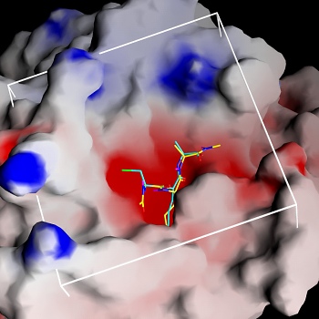
The docked arrangement of Ac-Ala-Tyr-Ala-Me bound to trypsin D189A,S190A is given in figure 4.2b. The ligand's position is very similar to the one observed in the docking against wt-trypsin (not shown).
The lowest energy structure found is consistent to the catalytic mechanism, with the carbonyl carbon situated next to the attacking S195 and the carbonyl oxygen positioned in the oxanion whole (S195 and G193). Clearly, the Tyr sidechain is not pointing to A190. Its OH-group, by contrast, seems to interact with the backbone oxygens both of G219 and (less obvious) A190.
The ligand surface is colored in accordance to the local interaction energy. The most evident interaction is calculated for the Tyr side chain's aromatic carbons.

Fig. 4.2b: Docking of a Tyr-containing peptide (blue) to trypsin D189A,S190A. Projected onto the van-der-Waals surface is each ligand atom's contribution to the interaction (red: neg. interaction energy, i.e. attraction, blue: positive interaction energy, i.e. repulsion). Protein residues interacting with the ligand are drawn in yellow.

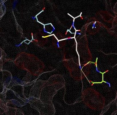
The next two figures are based on the same docking result. This time, the ligand is rendered with a solid surface. Again, the surface is colored according to the underlying ligand atom's interaction energy. In contrast to figure 4.2.b however, electrostatic and van-der-Waals interaction terms are displayed seperately.
Click the images to view an enlarged version.

Electrostatic interaction of AYA and Trypsin D189A,S190A Van-der-Waals interaction of AYA and Trypsin D189A,S190A
Fig. 4.2c: Electrostatic (left) and van-der-Waals (right) interaction of AYA and Trypsin D189A,S190A. The surface color follows the local interaction energy (red: negative, blue: positive). Selected protein carbons are drawn yellow, atoms especially contributing to the interaction are marked with "!".

Considering the above figure, the experimental findings can be explained: Binding of the Tyr side chain is not adversely affected by the mutation S190A, because the hydroxy group of Tyr can electrostatically interact with backbone oxygens of G219 and A(S)190. On the contrary, removing Oy of S190, in fact, promotes binding since this atom apparently "obstructs" van-der-Waals interaction between S190's Cß and aromatic ligand atoms.
As highlighted by the right figure, overall binding is mainly dominated by relatively unspecific van-der-Waals attraction between ligand atoms and the protein backbone.

4.3 Conclusion

The scope of enzymatic peptid ligations with the substrate mimetic approach is limited by the proteolytic activity of the enzyme. Detailed knowledge about the structure and mechanism of the serine protease trypsin facilitated the design of two protein variants addressing this problem. After removing the specifity-bearing side chains of both Asp 189 and Ser 190 the protease was successfully employed for ligating Arg- and Lys-containing peptides. Acyl transfer experiments with Tyr-containing substrates, however, revealed a raised specifity towards this amino acid. This is in agreement with docking simulations suggesting several van-der-Waals contacts and few electrostatic interactions with the protein backbone. Hence, the observed chymotrypsin-like specifity seems to be an intrinsic property of the S1 binding pocket.
One approach to overcome this limitation might be the introduction of a new specifity, favouring a substrate mimetic. The combined findings of experiment and modelling thus point to further design strategies and are a step towards a versatile man-tailored CN-ligating enzyme without any proteolytic activity. Have a look at our publication for a more detailed (and up to date) description of these results.

go to top
5. References
go to previous section  go to next section

Grünberg, R., I. Domgall, R. Günther, H.-J. Hofmann, and F. Bordusa (2000): Peptide Bond Formation Mediated by Substrate Mimetics: Structure-guided Optimisation of Trypsin for Synthesis. Eur J Biochem 267(24), 7024-30.

Thorman, M., Thust, S., Hofmann, H. J., Bordusa, F.(1999): Protease-Catalyzed Hydrolysis of Substrate Mimetics (Inverse Substrates): A New Approach Reveals a New Mechanism. Biochemistry 38, 6056-6062

Morris, G. M., Goodsell D. S., Halliday R. S., Huey, R., Hart, W. E., Belew, R. K., Olson, A. J. (1998): Automated Docking Using a Lamarckian Genetic Algorithm and an Empirical Binding Free Energy Function. J. Comput. Chem. 19, 1639-1662

last updated: 15/06/01

webmaster